Urology

Urology Treatments

Urethral Pathology and Urethra Stenosis

"Urethral pathology is more frequent than is believed and, if it is not treated in a timely and adequate manner, it can seriously jeopardize the integrity of the renal tissues and other genitourinary functions."

Play Video

Urethral Pathology and Urethra Stenosis

Table of Content

Play Video

Definiction

What is urethral pathology and urethral stricture?

The male urethra can suffer from a host of diseases. Congenital and infectious are treated in other chapters and tumorous are included in penile tumors. Therefore, we are going to focus on the description of urethral stricture, its causes, diagnosis and treatment, in order to give the reader a homogeneous content of this common pathology, whose correct diagnosis and approach from the beginning it will determine the subsequent evolution and the possibilities of successful treatment.

Stenosis of the urethra is a disease of medium prevalence that significantly alters the quality of life of the sufferer. Therefore, the fundamental message will be the adequate indication of the surgical technique to be used, avoiding starting with those (eg, internal urethrotomy) that are not simple because they are free of complications that can jeopardize the subsequent success of a correct approach.

Stenosis of the urethra is a disease of medium prevalence that significantly alters the quality of life of the sufferer. Therefore, the fundamental message will be the adequate indication of the surgical technique to be used, avoiding starting with those (eg, internal urethrotomy) that are not simple because they are free of complications that can jeopardize the subsequent success of a correct approach.

The narrowing of the urethra is defined as the marked decrease in the caliber of the lumen (or the canal) of the urinary urethra, associated or not with fibrosis in the adjacent tissues such as the prostate in the posterior portion or in the corpus spongiosum of the penis in the anterior portion of the urethra.

As the signs and symptoms of urethral stricture or narrowing can be very similar to those produced by both upper and lower urinary tract infections (UTI), or pathologies of the prostate (cancer, benign hyperplasia, etc.), it is possible that underdiagnosis if correct methods are not used to detect and measure it.

Who should I go to if I have urethral stricture?

Urologists play a fundamental role in the evaluation and treatment of urethral stricture since they are the medical specialists who are best familiar with the diagnostic techniques and the different therapeutic options directed to each particular case.

Causes of urethral stricture

The causes of this narrowing can be from:

Of all of them, inflammatory and infectious causes are the most common with 90% of all cases. However, traumatic causes are also frequent, especially in men who practice high-impact sports or who work in work environments lifting loads.

Iatrogenic causes are becoming rare and increasingly easily preventable.

In cases where it is impossible to know the cause of the narrowing of the urethra, the doctor qualifies it as an idiopathic stricture and it is treated as such, beginning with diagnostic tests.

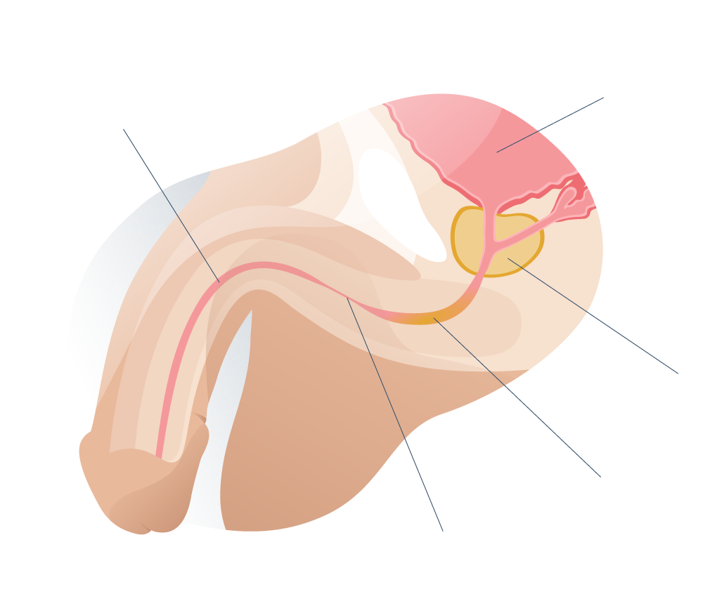
Anatomy of the urethra

The urethra is the only tube that carries urine from the bladder (where it is stored after being produced in the kidneys) to the meatus of the penis, where it can be eliminated.

In men, the urethra is not only longer (compared to the female urethra) but, for its medical study, it is divided into six segments, which are:


1 Bladder neck, at the base of the urinary bladder. This is the anatomical point in the urethra where the sphincter muscle prevents urine from leaving the bladder (urinary incontinence)..

2 Prostatic urethra, surrounded by the prostate gland. This segment is especially susceptible to obstructions due to inflammation of the prostate (cancer, benign hyperplasia, infectious or traumatic prostatitis, etc.).

3 Membranous urethra, from the lower edge of the prostate to the point where the urethra enters the bulb or root of the penis. This segment collaborates actively (or voluntarily) in the containment of urine.

4 Bulbar urethra, which begins when the urethra penetrates the root of the penis into the corpus spongiosum of the member. In this segment, the urethra is surrounded by muscle fibers of the bulbospongiosus which helps in the containment of urine voluntarily.

5 Penile urethra, from the base of the penis to the beginning of the navicular fossa on the penis. It is the most mobile segment of the urethra since the rest is very internal to bend or flex with the postures and the contour of the virile member.

Navicular fossa, a small dilation of the urethra (about one centimeter) inside the glans of the penis.

7 Meatus urethral, the most distal part of the urethra at the tip of the penis. It is linear in shape and measures about 5 millimeters.

Diferences between the male and female urethra

In the adult male, the urethra is usually about 8 inches long and passes through several important structures for sexual and urinary function. In women, the urethra is much shorter (approximately 3.5 cm) and empties into the vaginal introitus.

For this reason, urinary tract infections (UTIs) are much more common in female and male patients, because pathogens from the vagina can more easily ascend to the urethra and bladder. In severe cases, the infection can invade the kidneys and cause serious complications if not treated in time.

By consensus, the urethra is divided into two areas, and each of them into three others:

Anterior urethra: navicular fossa, penile and pendulous urethra and bulbar urethra.

Posterior urethra: membranous urethra, prostatic urethra and bladder neck.

In this chapter we will not deal with abnormalities of the prostatic urethra or bladder neck.

Looking for prices and information?

Use our virtual assistant and receive in your email everything related to the treatments that interest you. Budgets, recovery times, hospitalization, financing possibilities month by month, etc.

In just a few minutes and without any commitment.

En cumplimiento del Reglamento General de Protección de Datos le informamos que los datos por Vd. proporcionados serán objeto de tratamiento por parte de Andromedi con la finalidad de prestarle el servicio solicitado y/o contratado. Los datos no se cederán a terceros salvo en los casos en que exista una obligación legal. Para más información puede consultarla pinchando aquí.

Frequent pathologies of the urethra

Urethritis (gonococcal and non-gonococcal), hypospadias and lichen sclerosus can be found among the urethral pathologies that most afflict patients in urological consultation in addition to the so frequent urethral stricture.

Uretritis

A urethritis is an inflammation of the urethra, generally caused by pathogens and hence its conventional classification as gonococcal urethritis caused by neisseria gonorrhoeae, the bacteria that causes gonorrhea, or non-gonococcal urethritis caused almost always by chlamydia trachomatis (in the 20th 50% of cases).

The symptoms of urethritis, whatever its etiology, are characterized by manifestations of urinary obstruction such as pain and burning when urinating, urethral discharge of pus (especially in gonococcal urethritis) and a feeling of not having completely urinated.

Urethritis usually occurs in people who have had sex without a condom, but this is not always the case. Often times, inflammations of the urethra can occur from bumps (traumatic urethritis), tight underwear, irritating soaps, or after a season with a urinary catheter (in hospitalized patients, for example).

The diagnosis of urethritis begins with a medical history, questioning the patient (knowing the symptoms of it) and physical examination (to evaluate the signs). Laboratory tests such as analysis of urethral discharge may be indicated if this is the case or urinalysis (the presence of white blood cells in the urine is a sign of urinary infection).

Since the most common cause is infectious by bacteria, the treatment of urethritis consists of the administration of broad-spectrum antibiotics (AAE) and other antimicrobials, in addition to sexual abstinence for at least 7 days after the start of drug treatment. (Having sex could not only worsen inflammation but also spread the pathogen.)

Hygiene of the genital area is essential to reduce the presence of bacteria, especially in women, where the urethra is usually hidden inside the vagina.

Hypospadias

Hypospadias are the second most common type of urogenital malformation in men, after cryptorchidism (also known as maldescent of the testicle) and is characterized by being a structural abnormality of the urethra in which the external opening of the urethra (or meatus ) is not located at the tip of the penis but at some point on the ventral aspect of the member.

In 90% of cases, hypospadias occur at the level of the glans frenulum and this is positive for repair surgeries, the only treatment that exists.

Proximal hypospadias are less common and occur when the urethral meatus is located at the base of the penis, near the scrotum.

Most often, with hypospadia, there is an underdeveloped foreskin that reveals the internal structures of the penis near the site of the abnormal urethral meatus.

The causes of hypospadias remain a mystery but it is known with certainty that they occur due to genetic alterations during fetal development and that they are often part of some congenital intersex condition (such as hermaphrodism) or hormonal imbalances (for example, when urinary tract tissues are insensitive to testosterone and masculinizing chemical stimuli).

The surgery performed in hypospadias aims to:

In severe cases of hypospadia, skin implants and mucosa grafts may be required to restore the anatomy of the urinary canal and ensure erectile and reproductive function.

Lichen sclerosus atrophicus

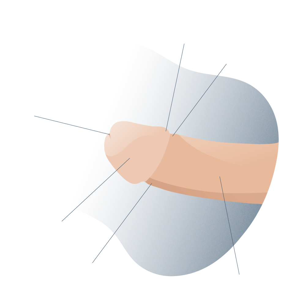
Anatomía general del pene

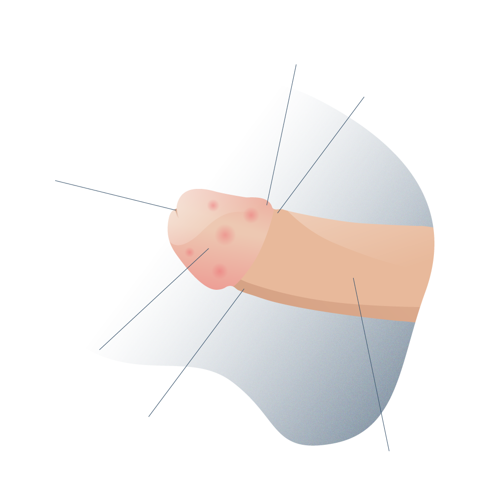
Healthy penis

Síntomas y signos de Balanitis

Penis with Balanitis

The Lichen sclerosus (name that is preferred to obliterating xerotic balanitis) is an autoimmune disease of unclear etiology, determined by a genetic predisposition (association with alopecia, vitiligo, diabetes and some autoimmune processes). It is also related to local aggression (radiotherapy, vulvectomy, sunburn, urine extravasation due to meatal stenosis, etc.) and may be related to an alteration of hormonal receptors and even infection. It is more frequent in women than in men and in these it predominantly affects the foreskin and glans. Lichen sclerosus has been linked to the development of squamous cell carcinoma of the penis.

Histologically, it is characterized by hyperkeratosis (thickening of the epidermis), parakeratosis, epithelial atrophy (tissue degeneration), hydropic degeneration of the basal lamina, lymphedema (or swelling), and a decrease in elastic fibers leading to loss of skin resistance.

Clinically, the skin and mucosa turn whitish, brittle, lose elasticity and often have small wounds, which are usually produced during sexual intercourse. This produces the development of phimosis and, in the case of urethral involvement, stricture.

It most frequently affects the foreskin, glans penis, urethral meatus and navicular fossa. When the involvement is limited to the foreskin, circumcision can be curative, although when there is already a stenosis of the meatus or navicular fossa, a surgical approach must be performed, which will be detailed later. An involvement of the more proximal urethra must always be ruled out, in order to adapt the type of intervention to the extent of the disease.

There is no definitive cure for lichen sclerosus but it is important for the patient to learn not to scratch with their nails, as it increases the likelihood of spreading the lesion or breaking the skin further. Wearing comfortable and loose clothing is also indicated.

Some topical creams or ointments bring relief from inflammation and itching, especially those that contain corticosteroids such as betamethasone or methylprednisolone furoate.

In some male patients, circumcision is indicated to reduce the area of contact with the lesion and reduce symptoms.

The best known complication of lichen sclerosus is the great risk of developing cancer if the treatment is not applied correctly, in the initial stages of the disease, which should be noted that it is not contagious.

Urethral stricture

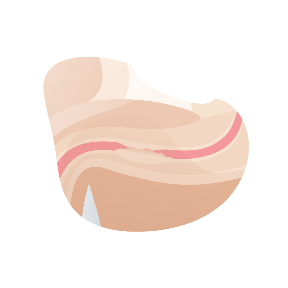
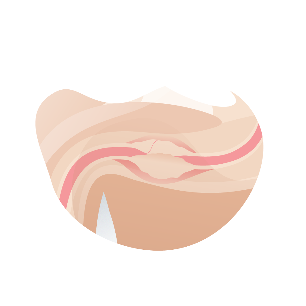
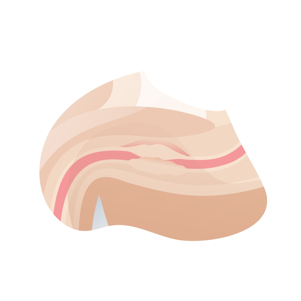
When we talk about urethral stricture, we do so referring to the anterior urethra, and it is a process that affects the urethral epithelium and the underlying spongy tissue. It consists of a scar fibrosis that reduces the diameter of the urethral lumen.

Any process that causes damage to the urethral epithelium or spongy tissue can cause stricture. One of the most frequent causes is traumatic. The incidence of lichen sclerosus is increasing and urethritis due to sexually transmitted diseases is no longer as common as a cause of urethral stricture.

Anatomia de una estenosis de uretra

Urethral stricture

How common is urethral stricture?

This is a urological pathology that has a high morbidity and that greatly affects the quality of life of patients.

It is estimated that its prevalence is close to 630 cases per 100,000 male patients around the world and the number of visits to the urologist for urethral stricture is 5,000 per year.

However, despite being a highly prevalent pathology, especially in men over 45 years of age, technological advances in diagnosis and treatment have significantly improved the prognosis and survival of patients.

It is also estimated that out of every 10 cases of urethral stricture, 4 of them take place in the bulbar segment of the urethra, at the base of the penis, given the large number of muscles in this area and the susceptibility to blows, especially in young patients or who play sports on a regular basis.

Doctor con estetoscopio

Diagnosis

Diagnosis of urethral stricture

Doctor con estetoscopio

The most common form of presentation is obstructive symptoms of the lower urinary tract, ranging from a decrease in the caliber of the voiding stream to acute or chronic urinary retention. It can also start with a urinary tract infection, prostatitis, or epididymitis. Frequently, the patient presents a decrease in the force of the jet progressively over a long time, to which he becomes accustomed. It is well tolerated until the appearance of some complication.

The suspected diagnosis is essential, especially in older patients, in whom the symptoms are attributed to prostate disease, although they do not improve with medical treatment, and the problem arises during access to the bladder during the intervention.

All male patients evaluated for obstructive symptoms in the urinary tract (whatever their type) should have a complete medical history in the to be investigated:

Some pathologies such as diabetes mellitus, certain cardiovascular diseases or habits such as smoking can significantly predispose to the appearance of exaggerated scar tissue on the walls of the urethra and lead to partial or total stenosis.

It is important to know the state of the erectile function of the penis and the quality of the ejaculations. Some men with partial urethral stricture may have difficulty conceiving a child.

To choose an appropriate technique for the surgical treatment of urethral stricture, it is necessary to know the following information regarding it:

The first three data can be known by one or more of the following techniques. However, the association of several of them is usually recommended to adequately plan the approach route and the surgical technique.

Ideally, it should be done with a thin rigid cystoscope, with a 0-30 ° optics, or with a flexible cystoscope. In addition to being able to perform an approximate calibration, the appearance of the urethral mucosa can be seen. The whiter it is, the more it points to a higher degree of spongiofibrosis.

Lo ideal es realizarla con un cistoscopio rígido fino, con óptica de 0-30° o con un cistoscopio flexible. Además de poder realizar un calibrado aproximado, se puede ver el aspecto de la mucosa uretral. Cuanto más blanquecina es, más apunta a un mayor grado de espongiofibrosis.

Es recomendable realizar esta técnica bajo la supervisión de un urólogo. En ese momento o previamente (con una sonda muy fina o a través de talla suprapúbica) se deberá haber llenado la vejiga de contraste. Consiste en la realización de radiografías en diferentes proyecciones oblicuas anteriores pélvicas previa introducción de contraste por uretra desde la fosa navicular de forma retrógrada. De ese modo, podremos obtener imágenes de llenado retrógrado, imágenes miccionales e, incluso, imágenes de uretrografía combinada con intento miccional a la vez del llenado retrógrado.

Se realiza con bujías de bola y es muy útil en estenosis distales. Tiene la limitación de que en caso de estenosis largas o múltiples no es muy valorable, ya que la más distal suele condicionar el estudio de las más proximales.

No se suele utilizar debido a que exigen un ecografista con experiencia y los resultados son habitualmente no concluyentes.

Una curva obstructiva con morfología «en meseta» apunta hacia una estenosis de uretra.
El grado de espongiofibrosis suele apreciarse realmente durante la intervención, aunque puede sospecharse de forma aproximada mediante el aspecto uretral durante la endoscopia, la mala distensión durante la misma, cuando se realiza la uretrografía retrógrada, por la existencia de manipulaciones o intervenciones previas y, eventualmente, en la ecografía.
Book your appointment in our center by clicking here

What is a urethroscopy and why is it so useful?

Currently, the gold standard in the diagnosis and evaluation of urethral strictures is urethroscopy associated with a cystoscope.

Urethroscopy is a minimally invasive diagnostic method that allows to quickly identify the exact location of the urethral stricture, its length, severity and the presence of other urethral pathologies such as:

Cystoscopy consists of raising the cannula with the video camera to the bladder, especially if the patient has associated pathologies of this organ due to urethral stricture.

What is urethroscopy?

In this diagnostic procedure, local anesthesia is used, generally, to avoid the discomfort caused by inserting a thin cannula into the urethra, especially if it is being compressed.

Once the anesthesia has taken effect, the urologist will insert the urethroscope into the urinary tract through the urinary meatus, at the tip of the penis and will ascend it until reaching the bladder, where he will release some 0.9% saline solution to distend the cavity and improve visualization (the latter if urethroscopy is accompanied by cystoscopy).

It can take up to 30 minutes for a complete urinary tract scan. There, the urologist or doctor in charge of the diagnosis will determine:

Urethral stenosis

Treatment Summary

We leave you here a summary of the approximate process from the appointment request, until receiving the medical discharge after the treatment. You can receive treatment at any of our centres in Madrid, Seville or Tenerife.

Necessary Consultations

At least two consultations will be necessary, one to collect information on the pathology and the other to perform a urethroscopy, a novel diagnostic method.

Hospital stay

Urethroplasty is a surgical procedure that requires the patient to remain hospitalized for a couple of days afterward.

Type of anesthesia

The anesthesia used is local but epidural anesthesia can often be used accompanied by anervous sedative.

Intervention Time

Urethroplasty lasts between an hour and an hour and a half but can often last less depending on the degree of tightness.

Postoperative

It is important that the patient rest for a week. There will be no large visible scars but it is important to eat well and rest.

Normal life

The normal life of the patient can be achieved practically instantaneously, the urinary function improves remarkably after a few days.

TECHNIQUES AND TREATMENTS

Treatment of urethral stricture

As a basis for treatment, it must be stated that there is no technique that serves to resolve any type or location of the stenosis, so a selection of the procedure should be made based on the following factors in relation to it:

1 Location of the stricture: that is, locate by means of a urethroscopy the exact site of the narrowing that is causing the problem.

2 Cause: the causes can be varied and it is essential to identify the factor that is causing the stricture in the urethra (traumatic, inflammatory, infectious, iatrogenic factors, etc.).

3 Length: the length of the urethral canal and the distance from the site of stricture to the urinary meatus on the penis can be used to identify the cause.

4 Number: generally, the urethral stricture is unique but it is possible to find two or even three points of narrowing. This complicates the treatment but it is necessary to repair all anatomical defects during surgery (even partial narrowing that may be beginning to appear).

· Tratamientos endouretrales ·

Localización

Localización

Causa

Causa

Longitud

Longitud

Número

Número

We have certain less invasive treatments for the urethra for the treatment of stenosis, which in many cases do not suppose a definitive solution to the problem, and even, if they are not indicated correctly, they can mortgage the subsequent treatment.

Types of urethral stricture

The urethra can be pathologically narrowed in different ways and to varying degrees, causing very characteristic signs and symptoms.

The main types of urethral stricture are:

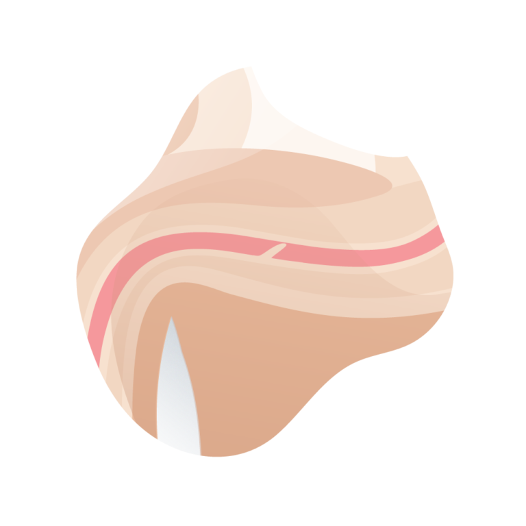
Mucous sheet

In the mucous fold, a thin layer of epithelial tissue develops that protrudes into the lumen of the urethral canal and causes a partial obstruction of urinary flow. This mucous fold is often short, but if it grows too large it can completely obstruct the urethra.

andromedi clasficacion de la estenosis de uretra 01

Mucous sheet

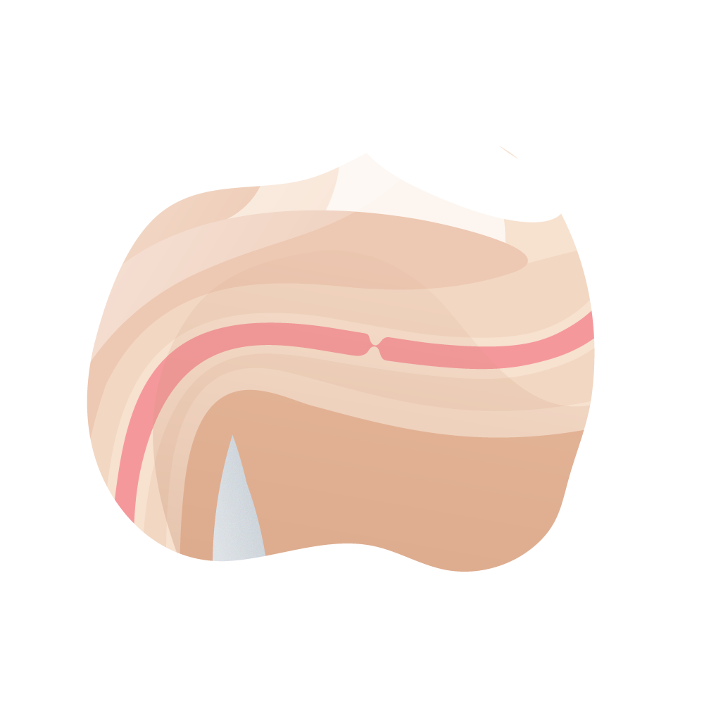
Espongiofibrosis

La espongiofibrosis es el crecimiento de tejido cicatricial en forma de un anillo delgado dentro de la uretra. La obstrucción puede ser parcial o total.

andromedi clasficacion de la estenosis de uretra 02

Espongiofibrosis

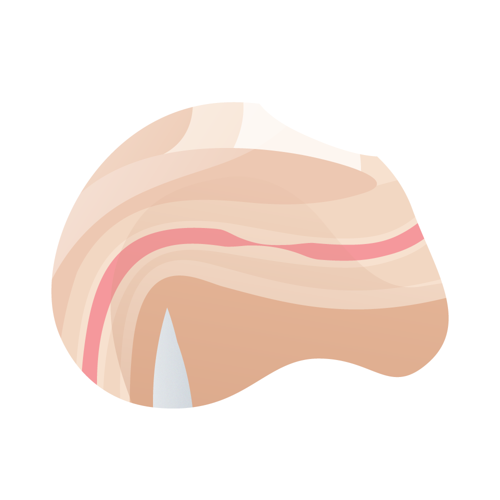
Iris stenosis

La estenosis en iris generalmente es más grande que la espongiofibrosis y el anillo (o estrechez) es más amplio. Se puede desarrollar lentamente y comprometer el flujo urinario o seminal de manera completa.

andromedi clasficacion de la estenosis de uretra 03

Iris stenosis

Spongiofibrosis and perispongium

In this case, the accumulation of scar tissue that obstructs the flow of urine or semen is not only urethral in nature but also from the spongy tissue of the penis. Treatment in this case is always surgical since the obstruction is usually total.

andromedi clasficacion de la estenosis de uretra 04

Spongiofibrosis and perispongium

Full thickness compromise, minimal spongy tissue fibrosis

This is one of the most severe types of urethral stricture that exists since the compression of the urethra is usually almost total. The inflammation is very marked and the urologist can tell when performing the urethroscopy.

andromedi clasficacion de la estenosis de uretra 06

Full thickness compromise, minimal spongy tissue fibrosis

Complete stenosis + fistula

As the name implies, men with this diagnosis have a completely obstructed urinary flow (representing a urological emergency that requires immediate surgery). The fistula is an abnormal communication when the pressure exerted by the accumulated urine in the bladder is very great, this defect must also be repaired surgically.

andromedi clasficacion de la estenosis de uretra 05

Complete stenosis + fistula

· Endourethral treatments ·

Urethral dilation

medico joven en operacion azul

It can be performed using more or less rigid instruments of progressively larger calibers, such as dilator plugs (safer in pendulous urethra), beniqués (for the bulbar and membranous urethra) or by means of a dilatation balloon inserted on a guide that, in turn , is placed under endoscopic control.

Dilation tears the stricture, not infrequently leading to a worsening of the stenosis as it is the cause of an increase in spongiofibrosis.

The best long-term indication for urethral dilation is membranous urethral stenosis after prostate surgery, since it is the only technique in which the external sphincter function is not at risk, thus preventing incontinence.

Periodic self-catheterization with pre-lubricated catheterization can also be used in patients with recalcitrant urethral strictures and difficult surgical solution, especially in elderly people or high surgical risk.

In all other cases, it is a temporary solution.

Internal urethrotomy

It consists of opening the narrowing of the urethra by making a transurethral incision. This implies the separation of the edges of the mucosa, allowing healing by second intention. A single incision may be made at 12 HC (in the most dorsal part of the urethra) or two lateral incisions at 10 HC and 2 HC.

The long-term studies carried out point to a failure rate of 58 to 84.3% and the results are worse in the penile urethra.

A higher rate of effectiveness of the technique has not been demonstrated due to the fact of repeating the procedure.

Therefore, the only reasonable indication for an internal urethrotomy would be a stricture with the following characteristics:

Thus, the chance of success is around 75%, which contrasts with 10-15% in the case of long, multiple or penile strictures, as well as in the case of a second internal urethrotomy.

Therefore, in the event of a failure of an internal urethrotomy, a urethroplasty should be performed.

operacion 1

Laser urethrotomy

It consists of the vaporization of the tissue that grows into the urethra. A type of laser that has little penetration into the underlying tissues should be used in order to avoid an increase in spongiofibrosis. Its results are so far similar to those of internal urethrotomy.

This type of surgical procedure is indicated for single urethral strictures, smaller than 2 cm regardless of its cause or location as long as the duct is not infected and the fibrosis of the tissue around the urethra (the same that causes the narrowing) is not very widespread.

Intraurethral tutors

They are placed endoscopically and should only be implanted in the bulbar urethra. They present frequent complications, such as postvoid drip, infection, irritative symptoms and intraurethral growth of fibrous tissue, which usually cause a new obstruction. This last complication is sometimes a problem that is difficult to solve, since the removal of the prosthesis is complex and, sometimes, requires excision of the treated urethral segment, which can lead to retraction of the urethra and a possible decrease in length penile.

It may be indicated in older patients with bulbar stenosis with little fibrosis and who have not been previously operated on.

andromedi tratamiento titulares 02

"In our centres in Madrid, Seville or the Canary Islands (Tenerife), we carry out the techniques and treatments with the most modern facilities and the best specialists and equipment to guarantee an effective solution".

· Urethroplasty ·

Urethroplasty with grafts and flaps

Two-stage urethroplasties (using scrotal flaps) and one-stage (using free preputial skin grafts) were the first major advances in urethral surgery, and prompted reconstructive surgery in the middle of the last century.

But these methods, which triumphed between the 1950s and 1970s, gave way to other techniques as a result of the incessant search for the ideal technique. Then the genital skin flaps in islets arose, with lateral, dorsal or ventral pedicles, based on an increasingly exact knowledge of the vascularization of the skin and fasciae of the penis and scrotum. First Orandi in England in 1968, then Hinderer in Spain in 1971 and later Standoli in Italy described various forms of genital skin islet flaps, which, once mobilized with their vascular connective pedicle, can reach all segments of the urethra to replace it totally or partially. The definitive popularization of island flaps belongs to Duckette in the field of pediatric surgery and to Quartey in that of adults.

Since then, in the last 20 years of the last century, the use of islet skin flaps constituted the basis of reconstructive surgery of the pendulous urethra and the anterior bulbar urethra. But, like all methods, urethroplasties with genital skin have limitations imposed precisely by the availability of donor areas. There are subjects who, due to being surrounded and / or having been operated on several times with poor results (urethral invalids), lack suitable skin for reconstruction. And it is also known that extragenital total skin free grafts do not provide predictable or reliable results. For this reason, regardless of the preputial mucosa graft (which is one of the best in urology), other grafts have been used, such as bladder mucosa and oral mucosa, which constitute the latest advance in the evolution of urethral reconstructive surgery. Both epithelia had been used more than 100 years ago in urology, but without achieving good results. More modern surgeons, with a more complete training, more refined technique and the possibility of antibiotic treatment were the ones who managed to consolidate them, first in the field of reconstructive surgery for congenital anomalies and later in that of urethral strictures.

The grafts can be classified according to their origin in:

The cutaneous or mucosal graft is a fragment of skin or mucosa that is totally disconnected from its location to be transferred to another remote place where it will develop a new vascularization.

The skin houses the cutaneous annexes, which include the sweat glands, sebaceous glands, hair follicles, and the erector hair muscle. These elements are practically absent in the skin and preputial mucosa and are one of the reasons that make it so suitable for urethral reconstructive surgery.

Once the graft is obtained and placed in contact with the bed where we want it to grow, it begins to adhere by means of the fibrin. It is important that, previously, all the fat and connective tissue adhering to the dermis have been removed, to facilitate graft survival.

During the first 3 days, the graft is nourished by plasma imbibition. Between the third and fifth day revascularization or inosculation begins. At 4 – 7 days the graft circulation, both blood and lymphatic, has been restored. Between the fourth and eighth day, epidermal proliferation is activated again and, in addition, there is a turnover of almost all collagen tissue. Tissue reinnervation, on the other hand, is slow (especially in elderly patients) and takes between 1 and 2 years.

The correct placement of the dressing, the use of pads or small cushions that compress the graft against its recipient tissue and the loose points that fix the central part of the graft to the underlying tissue (capitoné points) are very useful. During the first 5 days, the mobilization of patients must be, therefore, little to avoid the breakage of the very small newly formed capillaries.

Among the causes that facilitate graft necrosis, the most frequent is the formation of a seroma or hematoma between the graft and its bed. It can also be due to poor graft immobilization and the third cause of necrosis is infection.

Full-thickness skin grafts, which include the entire dermis and epidermis, can be used for urethroplasties. By including the reticular dermis, which contains a large amount of collagen and elastic fibers, it allows its extensibility and reduces the possibility of retraction. Likewise, it allows a better sensory reinnervation, which is recovered progressively during the first 2 years. Being very thick, it only ignites if the local conditions are optimal: absence of contamination and very well vascularized bed. If the donor skin has lymphedema (as occurs, for example, in patients with lichen sclerosus), the graft will have enlarged and malfunctioning lymph nodes, and will continue with lymphedema. They are grafts whose appearance is similar to normal skin, both in texture, pigmentation, presence of skin attachments and sensitivity. The usual donor sites for full-thickness grafts are the retroauricular, inguinal, and foreskin regions, as these are areas with thin skin thickness. They contract about 20%. In urology, the ideal grafts, both skin and mucosa, are foreskin. They are very elastic, they do not retract practically anything and they are very resistant to humidity. Once obtained, all the fat underlying the graft should be removed to facilitate its neovascularization.

The buccal mucosa has multiple advantages over the bladder: ease of obtaining, less morbidity, more manageability and shorter hospital stay. In addition, the success rate is higher because it has a thin lamina propria and a great wealth of vascular spaces in the submucosa, which facilitates the inoculation of the graft with new vessels. But within the enthusiasm it has aroused there are, however, disparate opinions. While there are authors who use these grafts at all levels of the anterior urethra (bulbar, penile, glandular, etc.), others prefer to use them only in the portion of the urethra that allows spongioplasty and coverage with the bulbocavernosa musculature. The results are so good at this level that the current trend is to consider oral mucosa free grafts the first option for treating bulbar urethral strictures (when excision and reanastomosis is not feasible). If, in addition, instead of grafting them on the ventral face of the urethra, the dorsal urethrotomy is used, first proposed by the Belgian Monseur in 1980 and later by Barbagli et al in 1995, the chances of success are maximized. This assertion is valid for both oral mucosa free grafts, as well as for those of bladder and preputial mucosa or penile skin.

In summary, as Jordan puts it, “In 40 years, urethral reconstruction methods have progressed from grafts to flaps and from grafts back to grafts. What has become clear during this time is that it is beneficial to have more than one option available to us and for surgeons undertaking urethral reconstruction they must master the full range of techniques available and understand the characteristics of substituent tissues.

· General indications for urethroplasties ·

The determining factors of the choice are multiple: age of the patient, general conditions of the patient, etiology of stenosis, location, number of stenosis (if multiple), degree of spongiofibrosis, length of the urethra, existence of complicating factors such as fistulas and intractable infection and the primary or secondary nature of the operation.

All of them have to be taken into account and, therefore, it is very difficult to make flow diagrams or simple and understandable algorithms.

The authors advise basing the choice, above all, on spongiofibrosis and location:

Very short, annular strictures

They occur at any level of the urethra in cases of mild internal urethrotomy fibrosis under vision.

Traumatic disorders of the membranous urethra

Bulboprostatic excision and reanastomosis.

Short strictures (<1 - 2 cm) of the bulbar urethra

Excision and reanastomosis (end-to-end urethroplasty).

Stenosis> 2 cm from bulbar urethra

Injerto de mucosa prepucial (oral en circuncidados):

  • Multiple, long strictures with great fibrosis and / or active infection in the anterior urethra.
  • Proximal half of the bulbar urethra. The graft is sutured ventrally, covered by a spongioplasty.
  • Distal bulbar urethra. Near the penoscrotal angle, it is preferable to suture the graft in the dorsal position, since the scarcity of urethral spongy tissue prevents a spongioplasty that completely covers the graft.
  • Two-stage urethroplasty, which can replace unusable portions of the urethra with grafts of preputial mucosa or, failing that, of oral mucosa or retroauricular skin. In cases of obliterative xerotic balanitis, it is important not to use preputial skin for reconstruction and to replace the last centimeters of the urethra completely with grafts of oral mucosa or retroauricular skin.
  • Uncomplicated penile urethral strictures
  • Transverse flap in islet of preputial mucosa. In circumcised patients, a distal penile circumferential skin flap or a ventral penile longitudinal skin flap can be used. In cases where the urethral stricture is not very closed, a urethroplasty with a dorsal graft of preputial mucosa or oral mucosa may be chosen.

· Graft types ·

For a century, the search for the ideal replacement for the male urethra has been relentless. It has not yet been found. However, at the present time, autologous epithelia of genital and extragenital origin allow urethral reconstructions in a single time with great reliability. Among all, the penile skin stands out (in particular, the preputial mucosa), either in the form of pedunculated islet flaps or in the form of free, full-thickness grafts; due to its versatility, wide availability, lack of hair, tolerance to humidity, flexibility and low tendency to shrinkage. Hence, the first option is still considered when it is available. Occasionally, however, there may be a deficit of penile skin, and then one must go elsewhere to obtain tissue. Long-term free full-thickness extragenital skin grafts give results that are far from satisfactory. That is why in complex situations free grafts of bladder mucosa or buccal mucosa have been used.

Preputial mucosa

Devine and Horton were the first to use foreskin grafts for hypospadias reconstruction. Kaplan later modified the technique. He used a tubular preputial graft to reconstruct the urethra and subsequently covered the ventral skin defect with a foreskin flap. He saw that there were no differences in the results between grafts and preputial flaps, always trying to orient the suture of the tubular plasties towards the albuginea.

Once the stenotic urethra has been opened until it has reached a healthy urethra, the length of the stenosis and its width are measured and the profile of the graft is drawn on the preputial mucosa extended and stretched over the albuginea of the penis. Given the elasticity of the preputial mucosa and in order to avoid oversizing the graft, its length should be equal to or slightly less than that of the stenosis. The width of the graft, which is oriented transversely over the foreskin, should be between 1.5 and 2 cm, although it depends on the severity of the urethral stricture. At this point, it is convenient to remember that the sum of the width of the stenosed urethra and that of the graft in millimeters is equal to the caliber on the Charriere scale of the future reconstructed urethra. The preputial mucosa incision should be shallow. It is then released from the subcutaneous tissue with scissors, trying to dissect the mucosa with as little underlying connective tissue as possible. (fig. 115 - 2). The preputial wound is closed with loose, short-term absorbable sutures without completing the circumcision, in case another preputial graft is required in the future. The graft is extended and then fixed with the help of insulin needles on a sterile splint. While an assistant irrigates the graft with saline and antibiotics to prevent desiccation, the surgeon removes the remnants of submucosal connective tissue adhering to the graft. At the end of bench preparation, the graft should be nearly transparent (Fig. 115-3). At this point, it is convenient to give 4 cardinal points of support to the graft, to facilitate its manipulation and transposition to the urethra. The graft is sutured with loose stitches to the urethra, using a 22 ch Foley catheter as a tutor. If the stricture is located in the middle or proximal bulbar urethra, it is preferable to suture the graft ventrally, since it can be completely covered by spongioplasty, which not only guarantees a good vascular bed, but also immobilizes the graft. The stitches include only the urethral mucosa and the graft, and a second plane of closure of the urethral cancellous must be given (spongioplasty). If the stenosis is of the distal bulbar urethra, we open the urethra dorsally and fix the graft to the albuginea of the corpora cavernosa according to the Barbagli technique. The albuginea provides a good bed to hold the graft and allows it to be immobilized extended, thus avoiding possible folds and blood collections that can make it difficult to inosculate the graft.

The points of urethroplasty include the urethral mucosa with its spongiosa on one side, and the cavernous albuginea and the graft on the other. Upon completion of the repair, the urethra is intimately attached to the corpora cavernosa. This technique should be applied with caution for pendulous urethral strictures in powerful men, as it can cause curving of the erect penis. To avoid this, very fine suture material (5/0) should be used and a small amount of albuginea should be caught with the stitches to avoid puckering or pleating it. On the other hand, if the graft is sutured ventrally in this portion of the urethra, the low thickness of the urethral cancellous prevents it from being sutured above the graft, having to resort to close subcutaneous flaps to cover the graft and provide adequate vascular supply to facilitate its neovascularization. It is for this reason that in the pendulous urethra it is preferred to use penile skin flaps rather than grafts; With the exception of the glandular urethra, where there is spongy to cover the graft.

Fistulas or strictures occur with foreskin grafts in 28% of cases (103 out of 368). Stock et al had 16% complications (44 tubular with 20% complications and 33 patches with 9% complications).

Bladder mucosa

The skin of the penis, particularly the mucosa of the foreskin, is free of hair, is thin, highly elastic and resistant to moisture; so it is still the favorite to rebuild the urethra. But when, as a result of unfortunate interventions, the entire skin of the penis has been consumed, or there is a scleroatrophic lichen (obliterating xerotic balanitis) that has rendered it useless, the reconstructive surgeon has to turn to other graft donation sites, usually extragenital skin full-thickness, hairless, following Horton and Devine. The results, however, are not always satisfactory when extragenital skin is used, due to the unpredictable, unpleasant and unacceptable contracture of the graft, which appears between 5 and 50% of cases. This led, as we discussed earlier, to the rediscovery of bladder mucosa grafts in the early 1980s.

Bladder mucosa is used if the abdomen must be opened at the same time to correct another problem or if the stricture to be repaired is very long and the oral mucosa does not reach. The danger in all these cases is that the grafts will fail due to the existence of abundant scar tissue. It is very important that there is no preoperative urine infection that could contaminate the graft. To avoid the problem of protrusion of the mucosa at the meatus, the most distal area of the urethra near the meatus can be reconstructed with preputial or penile skin. The bladder mucosa is used in reoperated cases with little available penile skin and no foreskin, although in these cases the oral mucosa is the first possibility if there is sufficient availability.

Info

Info

Technical aspects

The bladder approach is performed through a Pfannenstiel incision, the bladder is filled with serum (or 0.9% saline solution) and the detrusor muscle is incised until reaching the dark blue mucosa. A strip of bladder mucosa is then dissected, separating the detrusor to the sides. If the mucosa is accidentally opened, the dissection is done It is much more difficult, as the bladder empties and the mucosa does not remain distended. Before incising the bladder mucosa, the graft to be taken is marked with support points, and then the graft is removed. In cases of complete replacement, the graft is tubulated over a probe with continuous suture in the central area and loose stitches at the ends, to facilitate spatulation and finishing of the ends. The graft suture is oriented toward the albuginea to reduce the possibility of fistulas and is sutured to the ends of the open urethra with spatulation and loose stitches. The graft is covered with well vascularized tissue.

From the point of view of their behavior, bladder mucosa grafts are similar to full-thickness grafts. They contract little and tend to vascularize well, although when used to reconstruct the urethra they tend to dilate easily as soon as there is some area of relative stenosis, forming pseudodiverticula with some frequency.

Meatal stenosis and proliferation of the bladder mucosa exposed to the outside are common problems with this technique, which have led to the proscription of the reconstruction of the glandular urethra with bladder mucosa. Currently, in those cases in which it is necessary to reconstruct up to the meatus, mixed grafts are used, with penile skin or buccal mucosa for the most distal section. The major drawback of using the bladder mucosa is, however, the need to open the abdomen to obtain the graft, which implies greater morbidity, risks and inconveniences for patients, who require postoperative analgesia and longer hospital stays. All of this, together with a complication rate of more than 30%, led to the continuation of the search for new tissues for replacement urethroplasties. Hence, in the late 1980s and early 1990s oral mucosa grafts were rediscovered, abandoned due to septic complications since they were popularized by Humby in 1941.


Oral or buccal mucosa

The oral or buccal mucosa is one of the favorite grafts of reconstructive urologists, second only to the preputial mucosa when available. The buccal mucosa has a stratified, non-keratinized squamous epithelium and a lamina propria 0.5 mm thick. The epithelial cells of the intermediate stratum spinosum and the superficial stratum are firmly attached to each other and make the epithelium quite impermeable to urine. The mucosa is richly vascularized, which facilitates its rapid turnover and good inosculation.

In about 25 days, the epithelium can fully regenerate. The lamina propria is rich in elastic fibers and allows its elasticity, necessary with changes in the volume of the cheek. Mucosal sensitivity depends on trigeminal sensory branches and motor innervation of the buccinator muscle of the facial nerve. The graft can be obtained from the inner portion of each cheek and from the inner portion of the upper and lower lips. The length of the inner cheek grafts are usually 6 × 2 cm and those of the lip 4 × 1.5 cm. The length of the cheek graft can be increased by extending the graft to the lip, and can reach up to 10 cm. The oral mucosa does not tend to elongate or contract after being excised from the oral cavity, which allows the measurement made in situ in the mouth to be maintained during its preparation.

Usually, the transverse diameter of the graft is not usually sufficient to make a tube and is often used as a patch or tile. In very long urethral strictures, almost the entire urethra with bladder mucosa can be reconstructed, using oral mucosa to reconstruct the last centimeters and the meatus, in order to avoid ectropion and metaplasia suffered by the bladder mucosa at the urethral meatus.

Info

Info

Technical aspects

The ideal intubation during anesthesia is the nasotracheal one; although the orotracheal, displacing the tube laterally, can also allow access to a cheek and lips. Antibiotic coverage should be as for contaminated intestinal surgery: amoxicillin + aminoglycoside + metronidazole. The mucosa of the cheek is thicker than that of the lip, but the result for urethroplasties is probably the same. To obtain cheek mucosa, it is better to put a mouth opener with a lingual retractor. For the lower lip it is very useful to give 2 points of traction at each end of the lip to adequately expose its inner part. If the graft is removed from the cheek, it is advisable to mark the Stenon's canal at the level of the upper second premolar with a dermographic pencil or nylon thread.

It is recommended to infiltrate with 1% lidocaine with 1: 200,000 adrenaline and wait 7 min before beginning the dissection. This is done with scissors, trying to avoid the extraction of fat and buccinator muscle. The defect is sewn up with short-term absorbable stitches of 4/0. When the graft is obtained from the lower lip, more care must be taken, since the mucosa is thinner and tears more easily.

A useful technique is to make only the longitudinal incisions and dissect the subepithelial space between the two incisions underneath with scissors, as if making a submucosal tunnel, leaving the graft caught by the mucosa at its ends until the dissection is complete. The lip defect can be left without suturing and in 3 weeks the healing is perfect. It is preferable to suture it whenever it is estimated that labial asymmetries will not occur. Once the graft is obtained, it is immersed in saline serum and the whole team changes gloves.

On the bench, the graft is fixed with insulin needles and all the submucosal tissue is removed. We also take advantage of this moment to regularize the edges and ends of the graft. As we have already indicated previously, the graft is usually used as a patch and when a tube is to be made, it must be supplemented with a second graft or wrapped in a spiral around a probe. The immobilization of the graft is essential, which is achieved with an adequate surgical technique, a good dressing and a correct urinary diversion. The urethral catheter must be fixed and movements in and out must be avoided, as well as leakage of perisonous urine, which can wrinkle the graft during the first days. In the first 48 hours postoperatively, bed rest is recommended, provided that the graft is located in the pendulous urethra. If the graft is sutured in the bulbar urethra, the patient can be mobilized the day after surgery. One of the advantages of the buccal mucosa over the bladder is that it does not generate problems in the urethral meatus such as metaplasia, encrustation and overgrowth. The greatest disadvantage of the oral mucosa is its relative scarcity.

Comparing oral mucosa urethroplasties as a whole with the other free grafts, it seems that the former provide better results and are exposed to fewer complications. The graft that fares the worst is that of the bladder mucosa, due to double or more urethrocutaneous fistulas and the problem of ectropion of the external meatus, which does not exist for other epithelia.

Extragenital skin graft

When oral mucosa cannot be used and there is not enough foreskin or penile skin, due to previous reconstructive surgeries or lichen sclerosus, extragenital skin can be used as a graft. It can be obtained from the inner side of the arm, inguinal area or behind the ear region.

As a general rule, grafts used to partially reconstruct the urethra, such as urethral floor or ceiling, patch or tile, perform better than when used to reconstruct the entire circumference of the urethra, tube-shaped. It is more difficult for the entire surface of the tube to be covered and to be in contact with well vascularized tissue to guarantee inosculation of the graft, with partial necrosis of the graft being more likely to occur. After benching the full-thickness graft, with fat and subepithelial connective tissue removed, the graft should be nearly transparent.

One of the most common indications for the use of such a graft is obliterative xerotic balanitis. In these cases, the affected distal urethra must be removed and rebuilt in 2 stages with buccal or bladder mucosa grafts, to prevent the disease from reproducing. When buccal or bladder mucosa cannot be used, the best alternative is extragenital skin. Relatively large grafts are required when the urethra needs to be reconstructed in its entire circumference, and although the ideal is to use mucosa, retroauricular skin grafts perform as well in the long term as mucosal grafts, although they are easier to obtain.

Mesh skin graft

It is a partial thickness skin graft obtained from the inner thighs, buttocks, abdomen or inner arms. Generally, the graft is meshed with a mechanical mesh at 1: 1.5. It is an ideal graft to reconstruct the scrotum, rather than the urethra, since the mesh gives it a wrinkled, almost physiological appearance.

These grafts tend to retract and must be greatly oversized. At 2 weeks the area is fully epithelialized and at 8 – 12 weeks the epithelial surface is resistant and stable. With regard to this type of graft, a series of precautions must be taken into account. It should only be used for 2-step techniques. In hypospadias surgery, the graft should not be placed directly against the albuginea, since it forms a body with the latter and when retracting it produces an erection ventral bend.

Subcutaneous tissue should always be interposed to serve as a bed and care must be taken that the graft is not thick. After fixing the graft with resorbable loose stitches, it should be covered with tulgraso, trying to introduce part of it into the neomata to avoid if nechias. Dry gauze and a slightly compressive elastic bandage in X are placed on top of the fat tulle. The patient remains in bed for 7 days and is prohibited from sitting for another 7 days. The dressing is changed in 5-6 days, once the initial neovascularization has been established.

· General factors that influence the final result of urethroplasties ·

Previous surgery is one of the factors that most conditions the result of urethroplasties, which are obviously better in untreated patients. They are also better in strictures than in hypospadias. Within stenoses, those of traumatic origin behave better, followed by inflammatory and ischemic (by instrumentation or catheterization). Regarding the location, the greater stability and the existence of well vascularized beds make the bulbar urethral stenosis progress better than the penile one. Younger patients do slightly better than older ones and, logically, patients with urinary tract infection have a higher risk of failure.

Regarding postoperative urinary diversion, it is recommended to keep the urethral guard for 3 weeks in all types of free grafts. An attempt at voiding urethrography can then be performed with the catheter in place, helping to slightly mobilize the balloon so that it does not rest on the bladder neck. If there is no contrast leak, the catheter is removed and the urethrogram is completed. If there is a leak, the catheter is kept for at least one more week. In the case of end-to-end urethroplasties, control can be performed two weeks after the intervention, as in pedunculated grafts.

Book your appointment in our center by clicking here

MEDICAL EVALUATION

Andrological aspects of urethral stricture and its treatment

Urethral stenosis, insofar as it can affect the portion located in the penis and that both its symptoms and its treatment can have andrological implications, should be evaluated and treated always thinking about these aspects. Its fundamental implications will be reviewed below, and are also discussed in the chapters on disorders of erection (see Chapter 62) and ejaculation (see Chaps. 80 and 82).

Voiding disorders

Urethral stricture, both in terms of its cause and its evolution and after treatment, can, in addition to manifesting a decrease in voiding flow, have associated urinary infections, irritative symptoms, urethral or bladder stones. The discomfort that all this generates, together with the psychological implications that patients may have, can negatively influence the sexual life of the patient.

Ejaculation disorders

Urethral stricture may cause difficulty in ejaculation, and the patient may present a significant decrease in the output power of the seminal fluid (asthenic ejaculation). This can occur whether the stricture is proximal or distal to the bulbar urethra. A painful ejaculation can also be manifested, usually a consequence of urinary infection, especially prostatitis.

Chico pensativo

Erection disorders

Not infrequently does the patient with a urethral stricture present with associated erectile dysfunction. This can have the same cause as stenosis, especially in the case of severe pelvic trauma affecting the erector nerves or penile vascularization. It can also be a consequence of the surgical intervention that aims to resolve the urethral stricture. In this sense, it is important, whenever possible, to avoid damaging the penile innervation (especially in stenosis of post-traumatic origin of the membranous urethra) and the vascularization of the penis, respecting, for example, the bulbar arteries whenever reasonably possible when releasing the bulbar urethra. proximal.

On the other hand, an excessive release of the urethra in order to attempt an end-to-end urethroplasty at all costs instead of resorting to grafts in a too long stenosis, can produce ventral penile curvature during erection by traction of the shortened urethra.

As well The use of free dorsal grafts in the pendulous urethra can cause penile curvature, especially in very young patients, since said graft remains closely attached to the corpora cavernosa. To avoid this, very fine suture material (5/0) should be used and a small amount of albuginea should be caught with the stitches to avoid puckering or pleating it.

Patients with advanced lichen sclerosus can progress to destruction of the glans penis and a buried penis due to progressive retraction of the penile skin.

Info

Info

Summary

Urethral stricture is a relatively frequent and potentially disabling entity that must be solved by adequately planning the technique and evaluating the best options based on its cause, type, location, and extent.

A hasty choice of an apparently simpler technique (for example, an internal urethrotomy in a young patient with a 1-cm bulbar urethral stricture with spongiofibrosis) may limit the outcome of a subsequent urethroplasty, which, if indicated, at the time of diagnosis, it would have a better chance of success. The continuous training of the urologist, as well as the creation of reconstructive urology units in urology services, can significantly help patients suffering from this pathology.

Psychological disorders

Urethral stricture can cause difficulty in urination or ejaculation, or urinary infections, and even coexist with erectile dysfunction of organic origin, especially in cases of post-traumatic stricture. All of this can continue to occur after an intervention or as a consequence of it. This, together with the frequent need for multiple interventions throughout life and not infrequently from childhood, the presence of urethral or suprapubic catheters in the short or long term, and aesthetic alterations of the genital area, may lead the patient to need support and even treatment from the psychological and even psychiatric point of view. These disorders can create a vicious cycle that, especially in the case of erectile dysfunction, makes its treatment much more complex.

Frequently questions

Frequently asked questions at the Andromedi centres in Madrid, Seville and Tenerife

What happens if an infectious urethritis is not treated?

Complications from infectious urethritis (since non-infections may clear up on their own within a few days) are usually rare but should be avoided.

Among these complications due to urethritis are:

• Inflammation of the penis.

• The formation of purulent abscesses on the walls of the urethra.

• Total obstruction of the urethra, endangering the kidneys.

• Lymphangitis of the penis (an obstruction of the lymphatic drainage of the limb that can lead to tissue necrosis and amputation).

• Kidney infections such as acute pyelonephritis, which can cause irreversible damage to the kidneys.

Infectious urethritis (especially non-gonococcal) can leave sequelae such as epididymitis or inflammation of the epididymis, permanent infertility, conjunctivitis, infections of the skin and mucous membranes of the body, conjunctivitis and abundant discharge of pus through the penis, in addition to pain and erectile dysfunction.

How common are urethritis?

Although it may seem a thing of the past, sexually transmitted infections (STIs) such as gonorrhea and chlamydia continue to have a significant prevalence today within the population.

The World Health Organization (WHO) estimates that about 1% of all people have or have contracted gonorrhea and, between 3 and 4% have suffered from chlamydia, especially in low-income countries and the most affected are young people ( individuals between 16 and 24 years of age).

Prevention is the key and the best method is the latex condom (or condom) used properly.

What is the appropriate age to perform a hypospadia surgical repair?

The ideal age for hypospadia repair surgery depends on the type of malformation. Mild ones can be done between 3 and 18 months of age. The longer ones can wait longer (when the child is five years old) as long as urinary function is not compromised or there is cryptorchidism.

Can urethral stricture affect male sexual and reproductive function?

Yes, urethral stricture, like any other obstruction of the urinary and genital tract, can lead to serious male fertility problems, especially when the narrowing is chronic, partial, and long-standing.

It is important to remember that the urethra is not only the conduit for urine from the bladder to the penis but also the channel through which semen is ejaculated during sexual intercourse and, in cases of urethral obstructions, the semen containing sperm could not coming out, coming out too small, or retrograde into the bladder (a condition known as retro ejaculation harrow).

The stricture of the urethra in its acute stages or of semi-complete narrowing can cause intense pain in the penis if the erection is reached, which obviously will be quite difficult not only for the member to become rigid but also to have a lasting erection.

In some cases, urethral stricture can retract the corpus spongiosum during erection and cause a pathological bend, making penetration difficult.

How common is post-traumatic urethral stricture?

They usually occur from strong blows to the genital or perianal area, strong enough to generate a visible bruise and swelling in the internal structures. Most of the patients with this diagnosis are children who, after falls, have violently impacted this area with hard objects or young adults who practice high-impact sports, such as boxing, mixed martial arts or skiing.

Trauma urethral strictures are usually relieved by applying ice to the crotch and should clear up after a few days. However, if the narrowing of the urethra does not allow urination, it is necessary to operate surgically or use a urinary catheter to empty the urine (at best).

Is it possible to have sex with urethral stricture?

No, and in fact it could complicate the clinical picture of urethral narrowing, especially due to inflammatory or infectious causes. The ideal is to go to the doctor to be thoroughly evaluated and receive appropriate treatment. By reducing the narrowing of the urethra (and the causes that produce it), the patient can return normally to his sexual life.

Al hacer click en “comenzar” usted declara haber leído y aceptado nuestras políticas de privacidad. El responsable de los datos que introduzcas es la Clínica Andromedi, sin cederlo nunca a terceros. El envío de comunicación privada, así como de newsletters es la finalidad de su almacenamiento en la base de datos de andromedi.com (UE). En cualquier momento puede limitar, recuperar y borrar su información, entérese cómo aquí.

13403